Vertebral Column Anatomy
This slide illustrates the structure of the human vertebral column, detailing its key regions and individual vertebrae.
Layout & Structure:
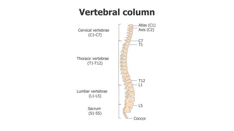
- The slide features a side view diagram of the vertebral column.
- It is divided into five main regions: Cervical, Thoracic, Lumbar, Sacrum, and Coccyx.
- Specific vertebrae are labeled, including Atlas (C1), Axis (C2), and the last vertebrae of each region (C7, T12, L5).
- The diagram clearly shows the natural curvature of the spine.
Style:
- The diagram uses a clean, illustrative style with clear labeling.
- The color palette is neutral, focusing on anatomical accuracy.
- The overall aesthetic is professional and educational.
Use Cases:
- Medical presentations and educational materials.
- Anatomy and physiology lessons.
- Explaining back pain or spinal injuries.
- Illustrating the relationship between the spine and nervous system.
- Patient education materials.
Key Features:
- Accurate anatomical representation.
- Clear and concise labeling.
- Visually appealing and easy to understand.
- Suitable for a wide range of medical and educational contexts.
Tags:
vertebral columnspineanatomymedicalbiologyskeletoncervicalthoraciclumbarsacrumcoccyxdiagrameducational
Ready to Get Started?
Impress your audience and streamline your workflow with GraphiSlides!
Install Free Add-onNo credit card required for free plan.