Heart Anatomy Diagram
This slide presents a detailed anatomical diagram of the human heart.
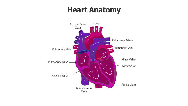
Layout & Structure: The slide features a centrally positioned, highly detailed illustration of the heart. Key components such as the atria, ventricles, valves, and major blood vessels (aorta, vena cava, pulmonary artery, pulmonary veins) are clearly labeled with connecting lines. The heart is depicted with a realistic, anatomical style.
Style: The diagram utilizes a color-coded scheme to differentiate between oxygenated and deoxygenated blood flow. The heart is rendered with a blend of purple and red hues, creating a visually appealing and informative presentation. The overall aesthetic is professional and medically accurate.
Use Cases:
- Medical education and training
- Presentations on cardiovascular health
- Patient education materials
- Anatomy and physiology lectures
- Illustrating blood flow through the heart
Key Features:
- Detailed and accurate anatomical illustration
- Clear labeling of key heart structures
- Visually appealing color scheme
- Suitable for a wide range of medical and educational presentations
- Professionally designed for clarity and impact
Tags:
Ready to Get Started?
Impress your audience and streamline your workflow with GraphiSlides!
Install Free Add-onNo credit card required for free plan.