Thyroid Gland Anatomy Illustration
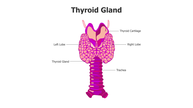
This slide presents a detailed anatomical illustration of the thyroid gland.
Layout & Structure: The slide features a central illustration of the thyroid gland, depicted with two lobes (left and right) connected by a thyroid cartilage. Below the gland is the trachea. Labels clearly identify the key components: Left Lobe, Right Lobe, Thyroid Gland, Thyroid Cartilage, and Trachea. The illustration is a single, focused element.
Style: The illustration employs a flat, vector-based style with a consistent purple color scheme. The design is clean and professional, prioritizing clarity and anatomical accuracy. There's a subtle use of shading to give a slight 3D effect to the gland and trachea.
Use Cases:
- Medical education and presentations
- Anatomy and physiology lessons
- Patient education materials
- Healthcare professional training
- Illustrating endocrine system components
Key Features:
- Clear and accurate anatomical depiction
- Professionally designed visual style
- Easy-to-understand labeling
- Suitable for a variety of medical contexts
- Visually appealing and informative
Tags:
Ready to Get Started?
Impress your audience and streamline your workflow with GraphiSlides!
Install Free Add-onNo credit card required for free plan.